Aftereffects
- Published13 Sep 2018
- Reviewed13 Sep 2018
- Source BrainFacts/SfN
Afterimages — what are they, and how are they generated by the color vision system of the brain?
This video is from the 2018 Brain Awareness Video Contest.
CONTENT PROVIDED BY
BrainFacts/SfN
Transcript
Let's try an illusion of the eye. at first sight this looks like a filtered picture of a person with a red dot on their nose but is there more to the image? let's find out. stare at the red dot on the picture for 20 seconds.
Now, blink at uniform color background, such as a wall. do you see what looks like a young woman who many of you may recognize as a singer ariana grande? maybe like this? Cool, right?
Why did you see what you saw when you viewed that image for some period of time? It's a phenomenon called after images. what are afterimages? afterimages are the reaction your brain has to a stimulus that saw before but no longer sees. to understand afterimages we must first understand the components of our optical system.
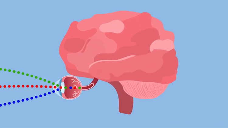
The Aqua pentalobe in our cerebral cortex is responsible for processing visual information. but where does this information come from?
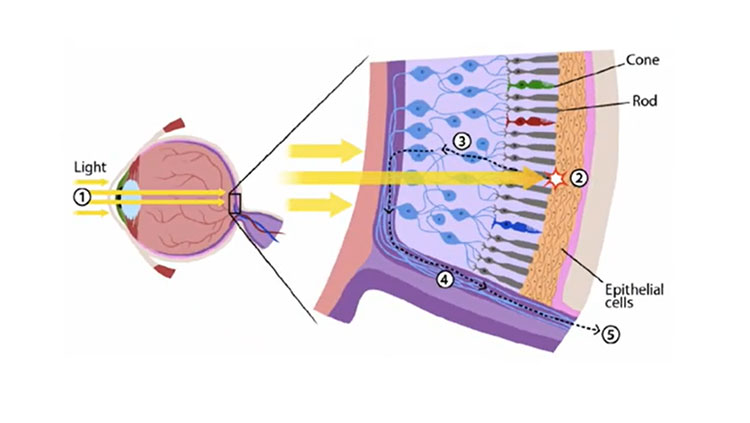
Our color perception is controlled by two main types of receptors in the eye. rods and cones. Rods and cones use phototransduction to convert the photons of light to enter our eye into electrical signals. then these electrical signals travel along the axis of our Chi seats, or retinal ganglion cells, which are our optic nerves. then optic nerves transmit the signal to the brain and then we see something. but individually, rods and cones control different aspects of our vision.
rod cells are extremely sensitive to light because it takes only a single photon to activate the photo pigment in them that sparks vision. this is why rods control our scotopic vision aka black and white night vision, since there is less light.
On the other hand, cones need a thousand photons to activate their photo pigment molecule, making them less sensitive to light. Therefore, cones control our photopic vision which is our color vision during daylight. near the center of our retina. the so-called fovea midget or small retinal ganglion cells, receive a signal from one cone while a thousand rods pass on one signal to parasol or large retinal ganglion cells in our peripheral vision area.
This is why our fovea gives such precise high equity images — also because there are three kinds of cones, each sensitive to a different color. These cells give us our color vision so since the solution has to do with colors playing with our mind, it is safe to say that cones are responsible for what we see.
How the cones work — when light hits a cone it undergoes a process where its structure has changed. this is called bleaching. this causes sodium and calcium ions as well as electrical currents to start flowing through the neural system, depending on how strong the light is. it may take a little while for the cone to return to its normal state when these cells in the retina are exposed to light for long periods of time. we see after images but why do we see different colors?
there are two different kinds of after images, positive and negative. in positive afterimages the afterimage is the same color as a stimulus.
However, negative afterimages show the complementary color to the color of the stimulus. the opponent-process theory explains how this occurs. the theory states that the human visual system interprets color using the difference between the activity of two sets of cones. for instance, the color red is seen when the cones that are most sensitive to long wavelengths. red light are more active than those that are sensitive to shorter wavelengths green light.
Now consider what happens when you look at bread for a while. the red cones get tired or bleached but the green cones are still fresh. now when you shift your eyes to a white background the green cones respond more than the tired red cones — which explains why the afterimage is green. so if exposed to one of the colors in the channel, there are three. the afterimage will be on the opposite color. a similar thing happens for the other two channels blue and yellow and black and white.
Let's go back to our original illusion and use all of this information to explain what we saw. we saw complementary colors to the original stimulus so we must have seen a negative afterimage. the blue tinge on her skin converted to a skin tone light color in the after image the green color on the lips and shirt showed up as a red lipstick and a red blouse.
Finally, the white coloring of her hair converts to a dark brown in the after image. put those components together and we get the image of ariana grande. knowing the signs behind the solution can also come really useful. next time you see this on Internet you can brag to your friends using the big words from this video and sound like a scientist because you know what's going on.
Also In Vision
Trending
Popular articles on BrainFacts.org


