C. elegans Have Special Multi-sensory, Multitasking Neurons for Movement
- Published9 Oct 2024
- Author Ailie McWhinnie
- Source BrainFacts/SfN
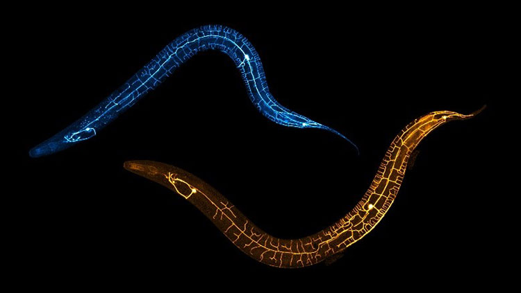
Neurons often specialize for specific tasks — some finely tuned to detect smells, while others respond to sound, light, or touch. But some neurons can process multiple senses simultaneously, like these PVD neurons pictured above in C. elegans.
C. elegans are tiny worms with a remarkably simple nervous system consisting of just 300 neurons. Their PVD neurons, glowing in gold and blue under the microscope, are responsible for sensing both harsh touch (like being poked) and proprioception, which is the ability to sense the body’s position in space.
Juggling the responses for two distinct senses poses a challenge for PVD neurons. When they detect a poke, they trigger the appropriate worm muscles to flinch away. But when they sense body movement, they relay feedback signals to muscles coordinating movement.
Unlike typical neurons that detect stimuli through their dendrites and send signals via their axons, PVD neurons do something different. The dendrites send proprioceptive signals to nearby muscles, while the axon is reserved for sending pain signals which trigger the flinch response.
Scientists at the National Brain Research Centre in India used a laser to selectively damage either the dendrites or the axons of PVD neurons and observed the worms’ movements.
With damaged dendrites, the worms still flinched, but their rhythmic movements became disjointed as their muscles failed to get necessary feedback. Damaged axons made the worms less responsive to being poked even though they moved normally, confirming the PVD neurons keep these two functions entirely separate. They do so by using different parts of same neuron for each sense.
Because C. elegans can regenerate axons and dendrites, the researchers observed the worms over 48 hours. After 24 hours, the damaged branches of the neurons were starting to heal and by 48 hours were almost fully repaired. By this time, the worms were moving and responding as normal again.
Humans can repair peripheral nerves, too, but only in early life and much more slowly than C. elegans. Because nerve repair is limited in adult humans, understanding the molecular pathways behind repair could one day allow nerve regeneration in nerve injury patients.
Study author Anindya Ghosh Roy emphasized the need to investigate both axon and dendrite repair. His team and others previously found dendrite repair isn’t realized using the axon repair pathway, but how it repairs itself is unknown. PVD neurons of C. elegans offer a powerful model to investigate this thanks to their large, accessible dendrites and the specific behavioral readout of dendrite function.
About the Author

Ailie is a PhD student in neuroscience at the University of Cambridge and a science writer with a particular interest in the intersection of life sciences with history, philosophy and society. Ailie was previously the Public Communications Intern at BrainFacts (Summer/Fall 2024) and editor-in-chief of EUSci and Women in Neuroscience UK.
CONTENT PROVIDED BY
BrainFacts/SfN
References
Brar et al. (2024). Functional Recovery Associated with Dendrite Regeneration in PVD Neuron of Caenorhabditis elegans. eNeuro, 11(5). https://doi.org/10.1523/ENEURO.0292-23.2024
Tao et al. (2019). Parallel Processing of Two Mechanosensory Modalities by a Single Neuron in C. Elegans. Developmental Cell, 51(5), 617-631. https://doi.org/10.1016/j.devcel.2019.10.008
Also In Movement


Trending
Popular articles on BrainFacts.org