Using Memory to Construct Representations
- Reviewed24 Apr 2023
- Author Marissa Fessenden
- Source BrainFacts/SfN

Your brain’s short-term memory storage is limited, so it builds simple constructs of people, places, objects, and events for reference. To really make sense of our moment-to-moment perceptions, the brain relies on its complex network of prior associations. These connections enable your brain to deal with variable perceptions. For example, you can identify a dog even if it is a different breed or color than any you have seen before. A bicycle still registers as a bicycle, even if it is obscured so that only one wheel is visible.
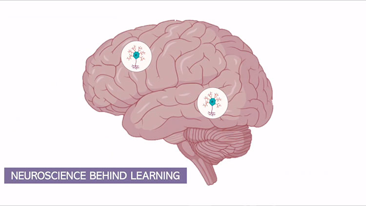
Constructing these representations relies on semantic memory, a form of declarative knowledge that includes general facts and data. Scientists are just beginning to understand the nature and organization of cortical areas involved in semantic memory, but it appears that specific cortical networks are specialized for processing certain types of information. Studies using functional brain imaging have revealed regions of the cortex that selectively process different categories of information such as animals, faces, tools, or words.
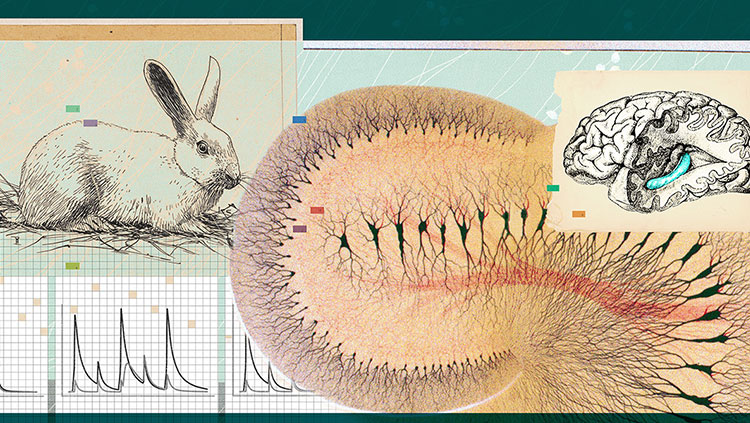
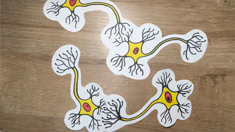
Recordings of individual brain cells’ electrical activity show that specific, single cells may fire when presented photographs of a particular person, but remain quiet when viewing photographs of other people, animals, or objects. So-called “concept cells” work together in assemblies. For example, the cells encoding the concepts of needle, thread, sewing, and button may be interconnected. Such cells, and their connections, form the basis of our semantic memory.
Concept cells reside in the temporal lobe, a brain area that specializes in object recognition. Scientists made great strides in understanding concept cells and memory by studying patient H.M. — a man who developed severe amnesia after undergoing an experimental surgery to quell his seizures. The seizures stopped and he could remember scenes, facts, and events that occurred before the surgery, but he could not form new memories. Researchers studying H.M. and others with similar deficits found that only patients who underwent tissue removal of certain areas of their medial temporal lobes — housing the hippocampus and adjacent regions we now know transform our transient perceptions and awareness into memories — experienced memory problems. Similarly, our understanding of thinking and language has been informed by studying people with unique deficits caused by particular patterns of brain damage.
Consider the case of D.B.O., a 72-year-old man who suffered multiple strokes. In tests run by researchers, D.B.O. could identify only one out of 20 different common objects by sight. He also struggled when he was asked to take a cup and fill it with water from the sink. He approached several different objects — a microwave, water pitcher, garbage can, and roll of paper towels — saying “This is a sink … Oh! This one could be a sink … This is also a sink,” before finally finding the real sink and filling the cup. But in striking contrast, he could easily identify objects when he closed his eyes and felt them; he could also name things that he heard, such as a rooster’s “cock-a-doodle-doo.”
Researchers concluded that D.B.O.’s strokes had damaged his brain in ways that prevented visual input from being conveyed to anterior temporal regions where semantic processing occurs. This blocked his access to the names of objects that he could see, but not his ability to name objects he could touch.
Adapted from the 8th edition of Brain Facts by Marissa Fessenden.
CONTENT PROVIDED BY
BrainFacts/SfN
References
Anaki, D., Kaufman, Y., Freedman, M., & Moscovitch, M. (2007). Associative (prosop)agnosia without (apparent) perceptual deficits: a case-study. Neuropsychologia, 45(8), 1658–1671. https://doi.org/10.1016/j.neuropsychologia.2007.01.003
Barense M. D., Warren. J. D., Bussey, T. J., Saksida, L. M. (2016) Oxford Textbook of Cognitive Neurology & Dementia, Chapter 4: The temporal lobes. https://academic.oup.com/book/24555/chapter-abstract/187755187?redirectedFrom=fulltext
Best, J. R., & Miller, P. H. (2010). A developmental perspective on executive function. Child development, 81(6), 1641–1660. https://doi.org/10.1111/j.1467-8624.2010.01499.x
Binder, J. R., Desai, R. H., Graves, W. W., & Conant, L. L. (2009). Where is the semantic system? A critical review and meta-analysis of 120 functional neuroimaging studies. Cerebral cortex (New York, N.Y.: 1991), 19(12), 2767–2796. https://doi.org/10.1093/cercor/bhp055
Florence Bouhali, F., Thiebaut de Schotten, M., Pinel, P., Poupon, C., Mangin, J. F., Dehaen, S., & Cohen, L. (2014). Anatomical Connections of the Visual Word Form Area. Journal of Neuroscience, 34(46) 15402-15414. https://doi.org/10.1523/JNEUROSCI.4918-13.2014
Buchsbaum, B. R., Hickok, G., Humphries, C. (2001). Role of left posterior superior temporal gyrus in phonological processing for speech perception and production. Cognitive Sci, 25, 663-678. http://www.sciencedirect.com/science/article/pii/S0364021301000489
Campbell, M. E., & Cunnington, R. (2017). More than an imitation game: Top-down modulation of the human mirror system. Neuroscience and Biobehavioral Reviews, 75, 195–202. https://doi.org/10.1016/j.neubiorev.2017.01.035
Centelles, L., Assaiante, C., Nazarian, B., Anton, J. L., & Schmitz, C. (2011). Recruitment of both the mirror and the mentalizing networks when observing social interactions depicted by point-lights: a neuroimaging study. PloS One, 6(1), e15749.https://doi.org/10.1371/journal.pone.0015749
Charpentier, C. J., De Neve, J. E., Li, X., Roiser, J. P., & Sharot, T. (2016). Models of Affective Decision Making: How Do Feelings Predict Choice? Psychological Science, 27(6), 763–775. https://doi.org/10.1177/0956797616634654
Dixon, M. L., & Christoff, K. (2014). The lateral prefrontal cortex and complex value-based learning and decision making. Neuroscience and Biobehavioral Reviews, 45, 9–18. https://doi.org/10.1016/j.neubiorev.2014.04.011
Domanski C. W. (2013). Mysterious "Monsieur Leborgne": The mystery of the famous patient in the history of neuropsychology is explained. Journal of the History of the Neurosciences, 22(1), 47–52. https://doi.org/10.1080/0964704X.2012.667528
Domenech, P. & Koechlin, E. (2014). Executive control and decision-making in the prefrontal cortex. Curr Opin Behav Sci, 1, 101-106.
http://www.sciencedirect.com/science/article/pii/S2352154614000278
Doré, B. P., Zerubavel, N., Ochsner, K. N. (2015). Social cognitive neuroscience: A review of core systems. In Mikulincer, M., Shaver, P. R., Borgida, E., & Bargh, J. A. (Eds.), APA Handbook of Personality and Social Psychology, Vol. 1. Attitudes and social cognition (pp. 693–720). American Psychological Association. https://doi.org/10.1037/14341-022
Frederick R. (2014). Testing for executive function in gibbons. Proceedings of the National Academy of Sciences of the United States of America, 111(13), 4738. https://doi.org/10.1073/pnas.1401589111
Hickok G. (2009). The functional neuroanatomy of language. Physics of Life Reviews, 6(3), 121–143. https://doi.org/10.1016/j.plrev.2009.06.001
Huth, A. G., de Heer, W. A., Griffiths, T. L., Theunissen, F. E., & Gallant, J. L. (2016). Natural speech reveals the semantic maps that tile human cerebral cortex. Nature, 532(7600), 453–458. https://doi.org/10.1038/nature17637
Konopka, G., & Roberts, T. F. (2016). Insights into the Neural and Genetic Basis of Vocal Communication. Cell, 164(6), 1269–1276. https://doi.org/10.1016/j.cell.2016.02.039
Mzuguchi N., Nakata, H., Kanosue, K. (2016) The right temporoparietal junction encodes efforts of others during action observation. Sci Reports, 6, 30274. https://www.nature.com/articles/srep30274
Peelle J. E. (2012). The hemispheric lateralization of speech processing depends on what "speech" is: a hierarchical perspective. Frontiers in Human Neuroscience, 6, 309. https://doi.org/10.3389/fnhum.2012.00309
Price C. J. (2012). A review and synthesis of the first 20 years of PET and fMRI studies of heard speech, spoken language and reading. NeuroImage, 62(2), 816–847. https://doi.org/10.1016/j.neuroimage.2012.04.062
Soutschek, A., Sauter, M., & Schubert, T. (2015). The Importance of the Lateral Prefrontal Cortex for Strategic Decision Making in the Prisoner's Dilemma. Cognitive, Affective & Behavioral Neuroscience, 15(4), 854–860. https://doi.org/10.3758/s13415-015-0372-5
Spunt, R. P., Satpute, A. B., & Lieberman, M. D. (2011). Identifying the what, why, and how of an observed action: an fMRI study of mentalizing and mechanizing during action observation. Journal of Cognitive Neuroscience, 23(1), 63–74. https://doi.org/10.1162/jocn.2010.21446
What to Read Next
Also In Learning & Memory
Trending
Popular articles on BrainFacts.org


