Early Experience Shapes the Brain for Life
- Reviewed25 Sep 2019
- Author Melissa Galinato
- Source BrainFacts/SfN

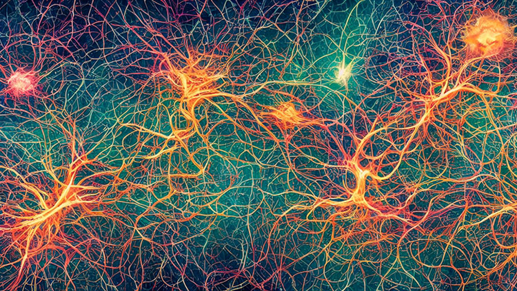
The brain’s ability to think, reason, and perceive all arises from an intricate web of connections among billions of cells. The connections that develop during infancy and childhood — periods of remarkable brain growth — can have lasting effects on learning and health.
Compared to other animals, we’re born with less developed brains, and they take longer to fully mature. Squirrel monkeys, for example, reach their adult brain size when they’re just six months old. One advantage of having such a protracted period of brain development is that our developing brains are more easily shaped by environment and experience, which helps us adapt and thrive in our unique environment.
The most sensitive windows of brain development are known as critical periods. During these times, sensory stimulation, motor activity, and even emotions shape the brain as it grows, wires, and learns. The period shortly after birth is one such window. A baby’s early life experiences — seeing parents’ faces, hearing their voices, and being held — provide important sensory information that guides its developing brain pathways.
Genes and environment both exert strong influences during critical periods. Together, they shape the neural circuits driving learning and behavior. One part of that process is forming new connections, or synapses, between neurons. Another is synaptic pruning, which removes weaker connections between cells. Even the death of some neurons helps the brain develop properly. Changes in neural connections during critical periods coincide with high rates of learning. Think of a baby learning to walk or a toddler learning language.
Understanding brain development is changing how we think about brain disorders and treatment. Some diseases emerging in teens and adults may have their roots in how the brain developed early in life. Schizophrenia, for example, is often thought of as an adult disorder because it typically emerges in the mid-20s. But newer research suggests it might arise because connections didn’t form properly in the developing brain. Other research suggests genes governing brain development could play a role in a person’s susceptibility to autism spectrum disorders. And, growing knowledge of how neurons form and wire up in young brains could guide treatments for brain injury. Once the stuff of science fiction, the idea of re-growing damaged neurons or pathways is getting closer to reality. Knowing how the brain was first constructed is an essential step toward understanding its later ability to reorganize and bounce back from injuries.
Adapted from the 8th edition of Brain Facts by Melissa Galinato.
CONTENT PROVIDED BY
BrainFacts/SfN
References
Dekaban, A. S. (1978). Changes in brain weights during the span of human life: Relation of brain weights to body heights and body weights. Annals of Neurology, 4(4), 345–356. doi: 10.1002/ana.410040410
Fu, M., & Zuo, Y. (2011). Experience-dependent structural plasticity in the cortex. Trends in Neurosciences, 34(4), 177–187. doi: 10.1016/j.tins.2011.02.001
Giedd, J. N. (2008). The teen brain: Insights from neuroimaging. The Journal of Adolescent Health: Official Publication of the Society for Adolescent Medicine, 42(4), 335–343. doi: 10.1016/j.jadohealth.2008.01.007
Giedd, J. N. (2015). Adolescent neuroscience of addiction: A new era. Developmental Cognitive Neuroscience, 16, 192–193. doi: 10.1016/j.dcn.2015.11.002
Holland, D., Chang, L., Ernst, T. M., Curran, M., Buchthal, S. D., Alicata, D., … Dale, A. M. (2014). Structural growth trajectories and rates of change in the first 3 months of infant brain development. JAMA Neurology, 71(10), 1266–1274. doi: 10.1001/jamaneurol.2014.1638
Leigh, S. R. (2004). Brain growth, life history, and cognition in primate and human evolution. American Journal of Primatology, 62(3), 139–164. doi: 10.1002/ajp.20012
Lenroot, R. K., & Giedd, J. N. (2006). Brain development in children and adolescents: Insights from anatomical magnetic resonance imaging. Neuroscience and Biobehavioral Reviews, 30(6), 718–729. doi: 10.1016/j.neubiorev.2006.06.001
Luciana, M., & Ewing, S. W. F. (2015). Introduction to the special issue: Substance use and the adolescent brain: Developmental impacts, interventions, and longitudinal outcomes. Developmental Cognitive Neuroscience, 16, 1–4. doi: 10.1016/j.dcn.2015.10.005
Shankle, W. R., Rafii, M. S., Landing, B. H., & Fallon, J. H. (1999). Approximate doubling of numbers of neurons in postnatal human cerebral cortex and in 35 specific cytoarchitectural areas from birth to 72 months. Pediatric and Developmental Pathology : The Official Journal of the Society for Pediatric Pathology and the Paediatric Pathology Society, 2(3), 244–259. doi: 10.1007/s100249900120
Sowell, E. R., Peterson, B. S., Thompson, P. M., Welcome, S. E., Henkenius, A. L., & Toga, A. W. (2003). Mapping cortical change across the human life span. Nature Neuroscience, 6(3), 309–315. doi: 10.1038/nn1008
Squeglia, L. M., Schweinsburg, A. D., Pulido, C., & Tapert, S. F. (2011). Adolescent Binge Drinking Linked to Abnormal Spatial Working Memory Brain Activation: Differential Gender Effects. Alcoholism, Clinical and Experimental Research, 35(10), 1831–1841. doi: 10.1111/j.1530-0277.2011.01527.x
What to Read Next
Also In Brain Development
Trending
Popular articles on BrainFacts.org


