Mapping the Way Forward for Brain Research
- Published26 Nov 2014
- Reviewed26 Nov 2014
- Author Mary Bates
- Source BrainFacts/SfN
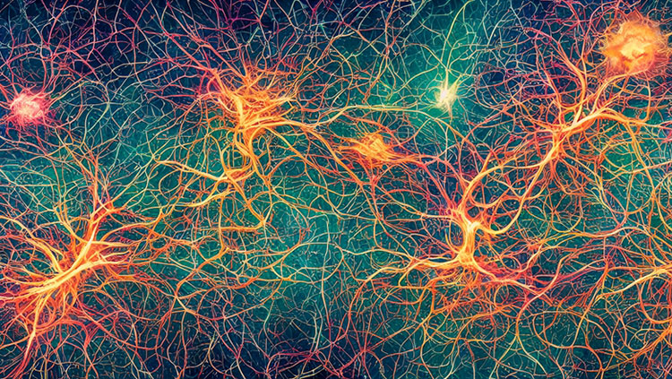
On any given day, the Beijing Subway shuttles millions of passengers between hundreds of stations. While impressive, it is nothing compared to the human brain, which transports innumerable signals between tens of billions of neurons.
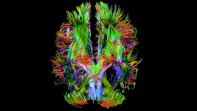
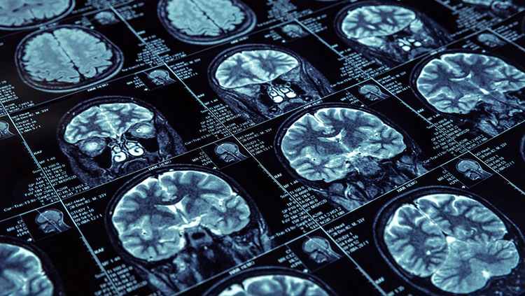
Human brain imaging techniques, such as diffusion tensor imaging (pictured above), cannot give scientists the same level of detail as the invasive structural and functional techniques used in animal studies can. However, efforts are currently underway to accelerate the development of new technologies capable of mapping and recording the interactions between individual cells and the circuits they form in various systems in the human brain.
To fully appreciate brain function, scientists need to know what’s going on in the various regions (or stations) in the brain, as well as how those regions connect (routes) and communicate with each other (schedule).
Whereas operators overseeing the Beijing Subway can track trains and traffic in real time, neuroscientists studying the brain lack a complete picture from which to pull such basic information. However, by studying animals such as rodents, which have simpler nervous systems and brains that are similar to those of humans, scientists have been able to obtain increasingly detailed information about the connections in the brain.
An atlas of the rodent brain
SfN Past President Larry Swanson played a major role in the early efforts to generate a map of the rodent brain. As a postdoctoral fellow in the 1970s, Swanson studied what goes on in the brains of rats when they are motivated to eat, drink, and have sex. He became determined to chart the connections of cells in the hypothalamus that were thought to be involved with these behaviors.
“Without knowing structural information, it’s hard to make sense of how the brain works and how to create a solution for motivation disorders like anorexia or overeating,” Swanson says. “At the time, the parts of the brain controlling these behaviors were a black box.”
To create a map, Swanson injected special tracers into the rat hypothalamus before carefully separating the brain into thin sections. Looking at the sections under a microscope, he then drew each cell and the connections between them by hand. This process took a long time and made it difficult for Swanson to share his results with others.
Then, in the 1980s, just as computer graphics were beginning to take off, Swanson realized that he could digitally capture the images that he saw under the microscope. By using a computer to stack these brain section images on top of one another, Swanson created the first 3-D digitized structural atlas of the rat brain. This brain atlas revolutionized the field, creating a standard brain map that researchers around the world could build upon.
“A brain map creates a database for all information about the brain,” Swanson said. “Once you have a map of the brain, you can add information about molecules, function, diseases, and more.
Gathering more details
Researchers are currently unable to capture the level of detail in the human brain that they have been able to collect in rodents. Major new research initiatives taking place around the globe have elevated the importance of understanding the human brain and unraveling the complexities of how it works. When President Barack Obama announced the Brain Research through Advancing Innovative Neurotechnologies (BRAIN) Initiative in April 2013, he set in motion a plan to accelerate the development of technologies to understand how “how individual brain cells and complex neural circuits interact at the speed of thought.” Neuroscientists realize that gathering such vast amounts of data will require new methods of data storage and analysis, and they are teaming up with experts in statistics, physics, mathematics, engineering, and computer science to make such methods a reality.
As some neuroscientists work on the creation of new technologies to probe the human brain, others are working on strategies to use detailed information about animal brain circuitry to enhance knowledge of normal and diseased human brain circuitry. To help reach this goal, Swanson and others are creating 3-D computer graphic models of the brains of humans and other animals that can interact and inform each other.
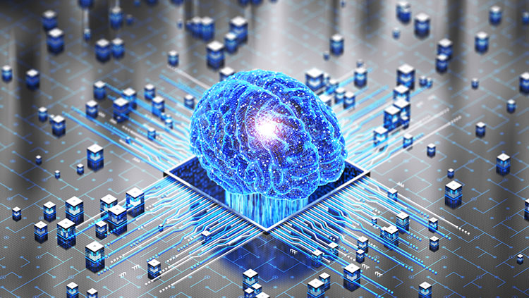
Ultimately, scientists aim to use data from maps and other new tools to understand how the brain functions to produce behaviors. To that end, scientists hope to build a computational working model of the human brain, which is a major emphasis of the European Union’s Human Brain Project.
“For a long time, research has been focused on how smaller and smaller chunks of the brain work,” Swanson says. “Now, scientists are trying to use the information about these pieces to put the brain back together to determine how this system works as a whole.
CONTENT PROVIDED BY
BrainFacts/SfN
Also In Tools & Techniques
Trending
Popular articles on BrainFacts.org


