Brainbows: Mixing Colors to Map the Brain
- Published6 Nov 2014
- Reviewed6 Nov 2014
- Source BrainFacts/SfN
Neuroscientists are creating a connectivity map of the brain to understand where neurons project and which neurons they’re talking to. But, just as it would be hard to find your way in a strange city without street signs, it’s difficult for scientists to map the brain when they can’t tell one neuron apart from its neighbors. This video from Vania Cao shows how scientists developed the “brainbow” technique to differentiate neurons by color, and it took third place in the 2014 Brain Awareness Video Contest.
CONTENT PROVIDED BY
BrainFacts/SfN
Transcript
To better understand our city, having a map of the roads and streets would help to identify important city structures and figure out how different parts of the city are connected. Based on the connections, we can then make educated guesses about the kinds of human activity that might happen in different areas.
Just like Google Maps for a city, neuroscientists have been working on a connectivity map for the brain. This “connectome” will help identify how neurons physically connect to one another.
This is no small task. If we take a satellite view of the earth, it’s hard to see where roads lead without visual labels. Similarly, it’s hard to tell one neuron with its many twisting branches, apart from its neighbors and their branches, because neurons are all the same color.
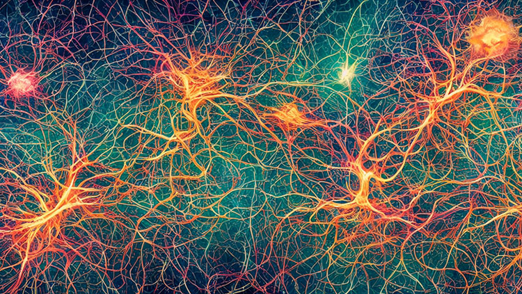
To shed some light on the situation, neuroscientists have come up with a way to brighten the brain with an artistic flair in mice! They do this by allowing individual neurons to make their own special mix of fluorescent protein “paint”, producing a rainbow-colored brain. This Brainbow technique lets us separate individual neurons by color. Think of it as color-coding neurons so we can easily see where they are, just like color coding transit lines on Google maps!
A Brainbow is made possible by what’s called “Cre-lox recombination”. Here’s a quick and dirty explanation of how the latest and greatest brainbow is made: We give neurons the genetic equivalent of a paint pot set; each pot contains the DNA recipe for a different color of fluorescent protein. This paint pot set has cutting sites between each pot of paint, called LoxP sites. A protein called Cre will randomly choose a pair of LoxP sites to cut, and will remove whatever paint pots are to the left of the cut. The neuron will only read the DNA recipe of the very next paint pot in what remains of the set. So, through the action of Cre, each paint set will produce only one randomly determined fluorescent protein “color”.
When we give each neuron many paint pot sets that each contribute a randomly determined color, every neuron will end up creating its own unique mixed color from the random ratios of their original fluorescent protein “paints”. These neurons are like tiny painters, mixing basic paint colors to create a new, complex shade. Now, we can tell almost 100 uniquely colored neurons apart in these beautiful Brainbows!
Brainbows are not just nice to look at; they differentiate neurons from their neighbors, and allow scientists to follow an individual neuron’s branches (the neuron’s axons and dendrites) as they criss-cross over other neuron’s branches and reach their final destinations and connection partners throughout the brain.
In the end, knowing where the subway lines in a city go won’t tell us how often the trains are running, or why they are on a particular schedule, or if the subway cars might break down or change direction. Likewise, knowing where a neuron is located and who it connects with, will not tell us everything there is to know about that cell’s function and role in producing thoughts, memories or disease. However, the Brainbow method of labeling neurons is a valuable tool that makes it possible to create detailed maps of neural connections, and provides a powerful, and beautiful, foundation with which to understand the basic structure of the brain.
Also In Tools & Techniques
Trending
Popular articles on BrainFacts.org


