Magnetic Resonance Spectroscopy
- Published1 Apr 2012
- Reviewed1 Apr 2012
- Source BrainFacts/SfN
Medical professionals have come to rely on even more specialized forms of the MRI machine, each suited to spotting specific warning signs.
Magnetic Resonance Spectroscopy
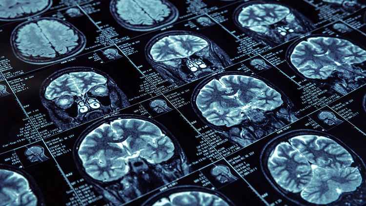
MRS, a technique related to MRI, uses the same machinery but measures the concentration of specific chemicals — such as neurotransmitters — in different parts of the brain instead of blood flow. MRS also holds great promise: By measuring the molecular and metabolic changes that occur in the brain, this technique has already provided new information about brain development and aging, Alzheimer’s disease, schizophrenia, autism, and stroke. Because it is noninvasive, MRS is ideal for studying the natural course of a disease or its response to treatment.
Functional Magnetic Resonance Imaging (fMRI)
One of the most popular neuroimaging techniques today is fMRI. This technique compares brain activity under resting and active conditions. It combines the high-spatial resolution, noninvasive imaging of brain anatomy offered by standard MRI with a strategy for detecting increases in blood oxygen levels when brain activity brings fresh blood to a particular area of the brain — a correlate of neuronal activity. This technique allows for more detailed maps of brain areas underlying human mental activities in health and disease. To date, fMRI has been applied to the study of various functions of the brain, ranging from primary sensory responses to cognitive activities. Given fMRI’s temporal and spatial resolution, as well as its noninvasive nature, this technique is often preferred for studies investigating dynamic cognitive and behavioral changes.
CONTENT PROVIDED BY
BrainFacts/SfN
Also In Tools & Techniques
Trending
Popular articles on BrainFacts.org


