How Does Social Isolation Affect the Brain?
- Published26 Nov 2024
- Author David Silverberg
- Source BrainFacts/SfN
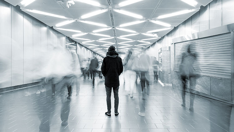
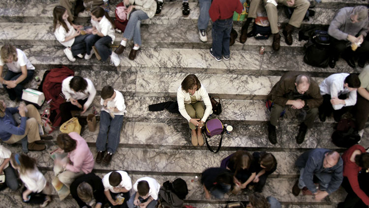
Rampant social isolation was among the most devastating outcomes of the COVID-19 pandemic. Amid school and workplace closures and stay-at-home orders, many people experienced an unprecedented challenge to their ability to maintain a healthy social life. While loneliness refers to not feeling connected to other people, social isolation involves not having access to other people to connect with.
Social isolation can take a significant toll on our mental and physical health. But researchers are still exploring how this experience plays out in the brain. Kay Tye, a neuroscientist at the Salk Institute for Biological Studies Systems Neurobiology Laboratory, is leveraging both mice and human studies to help explain how social creatures react to isolation.
Tye’s research explores the neural cells and structures associated with isolation. Her upcoming study, which is currently still under revision, focuses on how certain signals travel along various information highways — called anatomical projections — and how each of these paths “[gives rise to] different aspects of the emotional state we know as loneliness,” she explained.
Understanding how different parts of the brain respond to isolation gives researchers more opportunities to design new interventions and monitoring tools for those at risk of illnesses exacerbated by loneliness.
The Danger of Social Isolation
The social consequences of the pandemic were clear early in the crisis. An online survey conducted in 2020 by researchers at Harvard showed around a third of the roughly 950 respondents said they felt serious loneliness, which researchers defined as feeling lonely “frequently” or “almost all the time or all the time” in the month leading up to the poll.
Those numbers are significant because social isolation is associated with a long list of health challenges, including an increased risk of dementia, heart disease, and stroke. Socially disconnected people are also more likely to exercise infrequently, drink too much alcohol, smoke, and sleep poorly.

Tye’s research gives us a window into what our brains look like when influenced by isolation. She describes the anatomical projections of nerve cells as a constellation of circuits, each of which boasts different features. One projection contributes to the desire to explore the world around us. Another is vital for feeling averse to something, and the unpleasantness of being in that state. A different projection is important for boosting the motivation to seek social contact.
Tye seeks to dissect the tangled mess of wires within our brains to understand where each one goes, and how they contribute to our individual reactions to isolation. She believes those anatomical projections could be targeted to help people who are experiencing social disconnection.
If there were another global pandemic, for example, Tye wonders if it would be beneficial to help people keep the motivation to reconnect but lose the negative emotions associated with isolation, so they could maintain healthy social ties while avoiding the toll loneliness can take on health.
“That feeling of ‘I'm alone, and I want to seek social contact,’ there's nothing wrong with that,” she points out. “That’s adaptive. We want to keep that function, but we don’t need you to be so miserable and losing it while you’re doing that.”
How Prolonged Isolation Affects the Brain
Social connection is a basic human need. Surprisingly, in a human study, Tye found the desire to interact with others is similar to how the mechanisms in our body and brain can detect hunger. When that balance is thrown off, these systems aim to restore an equilibrium by signaling us to eat food or seek out relationships.
But social isolation can cause changes in the brain over time. In 2016, as an associate professor of brain and cognitive science at the Massachusetts Institute of Technology (MIT), Tye identified specific neurons in mice — dubbed dorsal raphe dopamine neurons — that light up during prolonged bouts of isolation. After 24 hours of isolation, those neurons become more sensitive to social stimulation, she said, compared to how they would behave in typical social situations.
Increased sensitivity to social stimulation doesn’t necessarily lend itself to successful social connection, though. Tye’s “social homeostasis theory” suggests that isolated creatures eventually stop trying to make new connections when their efforts consistently go unrewarded.
“At some point, we observe a drop in efforts being made. [People] stop calling, you stop going out, a mouse stops making vocalizations,” Tye said. “Your homeostatic social set point is being brought down to your new normal.”
Real-world Implications
Rebecca Saxe, a neuroscience professor at MIT who collaborated with Tye on her 2020 study on social cravings, remembers the research as a highly ambitious effort.
Forty participants spent a day in complete isolation. They then underwent three separate fMRI scans to assess how their brains responded to images of food and photos of people socializing with friends. Both images of food and socializing lit up the same areas of the brain — the substantia nigra pars compacta and ventral tegmental region.
When the study ended in January 2020, Saxe asked herself: Why would people be interested in the neuroscience of acute social isolation, especially among otherwise healthy and socially connected people? But when the pandemic hit in full force just two months later, she said, “the real-world relevance had become obvious.”
More than four years later, Tye is still digging into the neuroscience behind social isolation. A forthcoming study, currently under peer review, unpacks the differences between how male and female mice react to reintegrating with others after isolation. She found that females display a greater desire for social interaction than males when they’re reintegrated with peers after 28 days of isolation.
This data could be helpful in making different recommendations based on gender for people exiting quarantine.
Tye’s work, along with that of other researchers in her field, is filling the gaps in our understanding of social isolation and the brain, and the health care interventions that could help ease its most harmful consequences.
CONTENT PROVIDED BY
BrainFacts/SfN
References
Lee, C. R., Chen, A., & Tye, K. M. (2021). The neural circuitry of social homeostasis: Consequences of acute versus chronic social isolation. Cell, 184(6). https://doi.org/10.1016/j.cell.2021.02.028
Mathews, Gillian A., Tye, Kay. (2016). Dorsal raphe dopamine neurons represent the experience of social isolation. Cell. 164(4). 617-631. https://www.cell.com/cell/fulltext/S0092-8674(15)01704-3
Tomova, Livia, Tye, K., Saxe, R. (2020). Acute social isolation evokes midbrain craving responses similar to hunger. Nature. 23(12). 1597-1605. https://www.nature.com/articles/s41593-020-00742-z
What to Read Next
Also In Meet the Researcher
Trending
Popular articles on BrainFacts.org


