Glial cells help nerve cells communicate with one another by providing them with insulating layers of fats and protein called myelin, which allows the neuron’s electrical impulses to zip through axons without loss.
In the peripheral nervous system — where nerves in the body connect muscles and organs to the brain — these cells are called Schwann cells. When Schwann cells are altered, they often lose their ability to correctly perform their roles.
In a recent animal study in The Journal of Neuroscience, researchers at the University of Edinburgh examined the role of two specific proteins, periaxin and dystrophin-related protein 2 (Drp2), in Schwann cell growth, when myelination occurs.
Loss of one of these proteins is known to cause myelin damage in humans. In Charcot-Marie-Tooth disease, a relatively common hereditary condition, gene mutations and proteins compromise normal muscle functions, such as walking.
In this study, the scientists concluded not only are the proteins’ presence essential to proper Schwann cell growth, but so is their interaction.
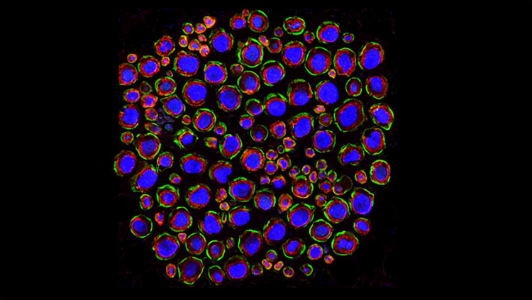
The image above shows a cross-section view of a mouse quadriceps nerve showing Drp2 staining in green in the plasma membrane of Schwann cells. Myelin is stained in red, surrounding the blue axons.
CONTENT PROVIDED BY
BrainFacts/SfN
Also In Cells & Circuits
Trending
Popular articles on BrainFacts.org


