Mapping the Human Connectome
- Published27 Oct 2017
- Reviewed27 Oct 2017
- Source BrainFacts/SfN
Creating a map of the most complicated terrain in the universe requires a host of special technologies.
Animation by Annie Campbell, Jim Stanis and Matt Wimsatt. Brainbow images provided by Jeff Lichtman, Harvard University. Diffusion tensor image provided by NICHD NIH.
CONTENT PROVIDED BY
BrainFacts/SfN
Transcript
In the early 1800s, Lewis and Clark set out to map the western United States. Charting the network of rivers that wound their way across the land.
Like those 19th century explorers, scientists have similarly been plotting the connections between the brain cells that underpin our every thought, action, and emotion. That effort began more than 100 years ago, when the Italian scientist Camillo Golgi formulated a way to stain individual brain cells called neurons. Neurons communicate with each other via chemical and electrical signals and Golgi’s technique enabled scientists to observe neurons in their entirety, including their specialized extensions, axons and dendrites, as they wound their way through brain tissue.
Golgi's discoveries ushered in a new era in neuroscience by giving scientists a glimpse of a key component of the nervous system. Even so the technique was limited: All neurons stained the same color and only a few could be visualized at a time. It was impossible to develop a comprehensive map or “connectome” of where and how each neuron’s dendrites connected with other neurons.
In the hundred years since, technologies Golgi couldn’t possibly have imagined are promising to deliver a “connectome” with remarkable resolution.
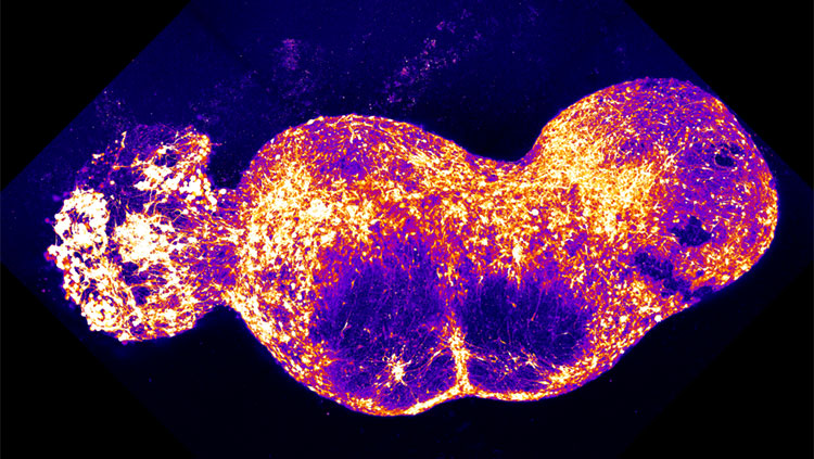
One of the most exciting developments uses a sophisticated genetic engineering technique to make neighboring neurons glow in a variety of colors.
Scientists introduce a random combination of four different fluorescent proteins into each neuron. As those proteins accumulate in varying amounts, roughly 100 different colors can emerge. The technique was dubbed Brainbow, appropriately enough, for the stunning rainbow-like images it creates. With each neuron receiving its own color, researchers can tease out the connections between them.
Brainbow is just one innovative technology scientists have employed to map connections in the brain. Even viruses can play a role. For example, when the rabies virus infects an animal or human, it attacks and spreads throughout the brain. By modifying the rabies virus to leave a trail of green fluorescent protein behind, researchers can map connections in the brain by tracking the virus as it jumps across the junctions between neurons known as synapses.
Fiber optics, too, have illuminated the workings of the brain. Using a combination of genetic engineering and fiber optics, scientists use flashes of light to activate neurons engineered to contain a light sensing protein. That activation stimulates the next neuron in the circuit. Scientists have already used the technology to examine disease-related circuitry in studies of Parkinson’s disease in a rodent.
Mapping the precise connections between individual neurons involves research with animals. New imaging technologies, however, are providing researchers tools to examine the human connectome.
For example, a noninvasive brain imaging technique called diffusion tensor imaging measures the behavior of water molecules in the brain. Water molecules tend to move along the length of nerve fibers, rather than side to side. Diffusion tensor imaging tracks the direction of water movement along the fibers allowing researchers to see the pathways connecting localized brain regions. By comparing diffusion imaging data from healthy and diseased people, researchers are learning how diseases and disorders affect brain circuits.
We can now look in a comprehensive way at which parts of the human brain are connected to each other. For the finer details of how neurons link up, we rely on information from experimental animals – at least for now.
Mapping the connectome is an enormous project, benefiting from the efforts of researchers throughout the neuroscience community. But just as it’s now possible to use Google Earth to track almost any route across the globe, we may one day know how any single neuron communicates with the rest of the brain.
Also In Tools & Techniques
Trending
Popular articles on BrainFacts.org


